Awake Intubation
Kristin Barkley, DNP, CRNA
Awake Intubation
- Awake tracheal intubation (ATI) is performed while the patient is spontaneously breathing, awake or lightly sedated.
- Techniques include flexible scope (nasal/oral) or video laryngoscope (oral).
- Nasal and oral approaches have similar success; the choice depends on clinician experience, equipment, anatomy, and bleeding risk.
- Indicated when intubation and/or ventilation are expected to be difficult (see Difficult Airway Algorithm).
- Goal: maintain airway reflexes and spontaneous ventilation throughout.
- Contraindications:
- Uncooperative patient.
- Airway bleeding or secretions obscuring the view.
- Local anesthetic allergy.
Procedure Guide
Planning
- Determine route (nasal/oral) and required equipment.
- Ensure backup airway plan and rescue equipment are available.
Equipment
- Nasal: RAE ETT (various sizes), nasal trumpets, lubricant.
- Supplemental oxygen via nasal cannula or high-flow nasal system.
- Video laryngoscope or flexible intubating scope (FIS).
- FIS with suction and irrigation/local ports; use anti-fog spray.
- Oral/nasal airways of multiple sizes.
- Suction setup.
Medications
- Antisialagogue: Glycopyrrolate 0.2 mg IV 15 minutes prior to reduce secretions.
- Topical nasal vasoconstrictors: Afrin or phenylephrine spray.
- Topical local anesthetic: cotton pledgets soaked in 4% cocaine or lidocaine with epinephrine for vasoconstriction and anesthesia.
- Technique: place along the upper border of the middle turbinate at a 45° angle to the hard palate, advancing posteriorly toward the sphenoid.
- Sedation (titrate slowly): dexmedetomidine, ketamine, midazolam, propofol, fentanyl, or remifentanil.
- Local anesthetic techniques:
- Nasal: sphenopalatine and anterior ethmoidal nerve blocks (nasal cavity, soft palate, tonsils).
- Oral: glossopharyngeal block or 2% viscous lidocaine gargle for gag suppression.
- Laryngeal: transtracheal and superior laryngeal nerve blocks.
- Nebulized 4% lidocaine may supplement topicalization.
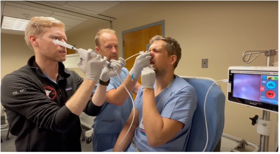
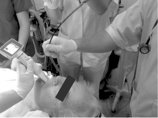
Awake Nasal Flexible Scope Intubation
- Operator may stand at the head or side of the bed (frontal approach acceptable).
- Load ETT onto the FIS.
- Advance to visualize the glottis; confirm tracheal entry and pass the ETT.
- Troubleshooting:
- Suction secretions before advancing ETT.
- Withdraw and clean lens if visibility is poor.
- Assistant may perform jaw thrust to improve view.
- If ETT hangs on arytenoids, rotate 90° counterclockwise and re-advance.
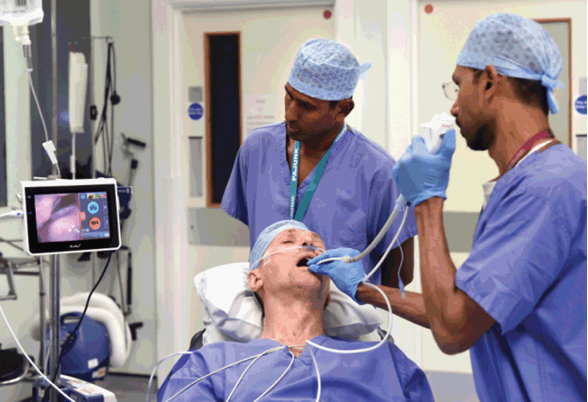
Awake Oral Video Laryngoscopy Intubation
- Use bite block if using FIS in conjunction.
- Patient in slight head elevation or sniffing position.
- Operator at head of bed for visualization and manipulation.
SCOPe Guide
Strategies
- Patient preparation: administer preoperative medications (antisialagogue, vasoconstrictors, topical anesthesia) with sufficient time for onset.
- Preoxygenation: semi-Fowler or upright position; continuous nasal or high-flow oxygen during the procedure.
- Equipment readiness: ensure all devices, suction, and medications are immediately available.
- Sedation: titrate slowly to preserve spontaneous ventilation and airway reflexes.
- Confirmation:
- Visualize tracheal rings beyond ETT tip before removing scope.
- Confirm placement with ETCO₂ over several consistent breaths.
- Induction: may proceed once airway is confirmed secure.
Pearls
- Have multiple ETT and airway sizes ready before starting.
- Optimize patient comfort and safety with adequate topical anesthesia and antisialagogue.
- Proceed slowly and deliberately—visualization and oxygenation take priority over speed.
- Suction secretions frequently and maintain lens clarity.
- Communicate continuously with the patient if awake or lightly sedated.
Media
References
- UpToDate: Flexible Scope Intubation for Anesthesia
- Elisha S, Heiner JS, Nagelhout JJ. Nurse Anesthesia. 7th ed. Elsevier; 2023.
Media Attributions
- Picture2
- Picture1
- Picture3
