Cardiac Function Assessment
Bailey Freeman, DNP, CRNA and Angela Mordecai, DNP, CRNA
Quick Facts
- Visual estimation accurately approximates formal measurements
- Both left and right ventricular function can be rapidly assessed
- Qualitative assessment is often sufficient for clinical decision-making
- Multiple views increase accuracy of functional assessment
Indications
Cardiac function assessment via POCUS is indicated for:
- Shock states of uncertain etiology
- Suspected heart failure
- Monitoring response to interventions
- Pre/post-procedure cardiac evaluation
- Risk stratification in acute presentations
Absolute Contraindications
- No specific contraindications beyond general POCUS limitations
Relative Contraindications
- None specific to cardiac function assessment
Procedure
Equipment Needed:
- Previous views obtained (parasternal, apical, subcostal)
- Clips recorded for dynamic evaluation
- Ultrasound machine with cardiac presets
- Image storage capacity for review
Functional Assessment Technique
LEFT VENTRICULAR FUNCTION
- Obtain and optimize standard views:
- Parasternal Long Axis (PLAX)
- Parasternal Short Axis (PSAX) at mid-papillary level
- Apical four-chamber view
- Identify end-diastolic frame (largest LV cavity size)
- Identify end-systolic frame (smallest LV cavity size)
- Compare cavity size reduction visually
- Evaluate wall motion in all visible segments
- Assess for uniform myocardial thickening during systole
- Classify function based on visual estimation
RIGHT VENTRICULAR FUNCTION
- Utilize apical four-chamber and subcostal views
- Compare RV size to LV (normally RV 2/3 or less of LV size)
- Assess RV free wall motion for thickening and excursion
- Evaluate tricuspid annular movement if visible
- Look for septal position and motion:
- Normal: Septum curves toward RV
- Abnormal: Flattening or bowing toward LV
- Assess RV systolic function qualitatively
COMPREHENSIVE ASSESSMENT
- Integrate findings from multiple views
- Assess both systolic and diastolic function when possible
- Evaluate structural abnormalities that may affect function
- Correlate with clinical presentation and vital signs
Function Assessment Details
LEFT VENTRICULAR SYSTOLIC FUNCTION
Assessment Methods
- Visual estimation:
- “Eyeballing” technique (most common in POCUS)
- Compare end-diastolic to end-systolic volumes
- Evaluate endocardial excursion
- Classification system:
- Normal: EF >55% (vigorous contraction)
- Mild dysfunction: EF 45-54% (reduced contraction)
- Moderate dysfunction: EF 30-44% (obviously reduced)
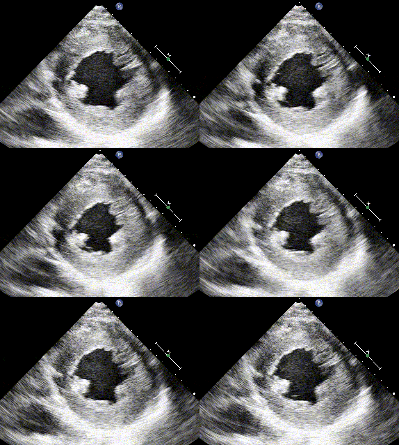
- Severe dysfunction: EF <30% (minimal contraction)
Wall Motion Assessment
- Regional abnormalities:
- Evaluate in multiple views
- Describe location using standard segments
- Classify as normal, hypokinetic, akinetic, or dyskinetic
- Global function:
- Overall contractility assessment
- Chamber size relationship to function
- Wall thickness evaluation
RIGHT VENTRICULAR FUNCTION
Assessment Approach
- Size and shape:
- Normal: RV ≤2/3 size of LV, triangular shape
- Dilated: RV >2/3 size of LV, rounded shape
- Severely dilated: RV equal to or larger than LV
- Function evaluation:
- Free wall motion and thickening
- TAPSE (Tricuspid Annular Plane Systolic Excursion) if measured
- Interventricular septal motion and position
Signs of RV Dysfunction
- Pressure overload:
- RV hypertrophy
- Septal flattening in systole
- D-shaped left ventricle in short axis
- Volume overload:
- RV dilation
- Septal flattening throughout cardiac cycle
- Reduced RV systolic function
DIASTOLIC FUNCTION CLUES
Limited POCUS Assessment
- Indirect signs:
- Left atrial enlargement
- LV hypertrophy
- IVC dilation with reduced respiratory variation
- Clinical correlation:
- Preserved EF with heart failure symptoms
- Signs of elevated filling pressures
- Note: formal diastolic assessment typically requires spectral Doppler
Confirmation Methods
MULTI-VIEW ASSESSMENT
- LV function confirmation:
- Assess in at least two different views
- PLAX and PSAX provide complementary information
- Apical view offers four-chamber perspective
- RV function confirmation:
- Apical and subcostal views most useful
- Both size and function assessment
- Septal position evaluated in multiple views
- Correlation across views:
- Consistent findings increase confidence
- Discrepancies require further evaluation
- Technical limitations should be noted
CLINICAL INTEGRATION
- Correlation with vital signs:
- Blood pressure
- Heart rate
- Respiratory status
- Physical examination findings:
- Jugular venous distention
- Pulmonary examination
- Peripheral edema
- Hemodynamic impact assessment:
- Evidence of end-organ perfusion
- Response to interventions
- Overall clinical picture
Documentation Requirements
- Qualitative assessment of ventricular function
- Chamber size relationships
- Wall motion abnormalities if present
- Integration with clinical presentation
- Views utilized for assessment
- Technical limitations encountered
- Relevant secondary findings (effusion, valve abnormalities)
SCOPE GUIDE
Strategies & Clinical Optimization
Visual Estimation Techniques
- Eyeballing approaches
- Identify true end-diastole and end-systole
- Compare cavity size reduction visually
- Look for approximately 50-60% reduction in cavity size
- Categorization process
- Normal: Vigorous contraction, >50% reduction in cavity size
- Mild dysfunction: Slightly reduced contraction, 40-50% reduction
- Moderate dysfunction: Obviously reduced, 20-40% reduction
- Severe dysfunction: Minimal contraction, <20% reduction
- Wall motion evaluation
- Assess thickening of myocardium during systole
- Look for regional variation in contraction
- Pay attention to segments with reduced motion
Right Ventricular Assessment
- Size comparison technique
- Compare RV:LV ratio in apical and subcostal views
- Normal RV is approximately 60% of LV size
- Dilated RV approaches or exceeds LV size
- Function assessment approaches
- Free wall motion evaluation
- Tricuspid annular movement tracking
- Septal motion and position analysis
- Overload pattern recognition
- Volume overload: Dilation with septal shift throughout cycle
- Pressure overload: Hypertrophy with systolic septal flattening
- Combined: Features of both patterns
Clinical Integration Strategies
- Shock differentiation
- Cardiogenic: Poor LV function with signs of congestion
- Distributive: Hyperdynamic LV with normal/small chambers
- Obstructive: RV dilation/dysfunction, pericardial effusion
- Hypovolemic: Small, hyperdynamic chambers, IVC collapse
- Serial assessment approach
- Documented baseline function
- Monitoring response to interventions
- Comparison to prior examinations
Pearls
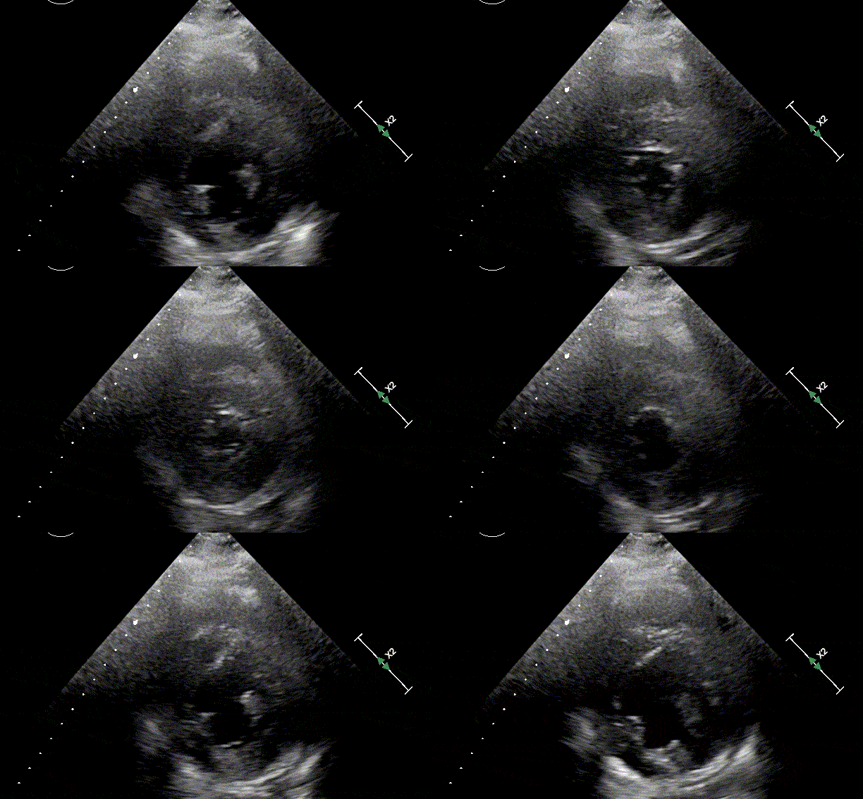
- A normal heart will reduce its cavity size by more than half during systole
- Septal flattening or bowing toward LV suggests RV pressure/volume overload
- Diffuse vs. regional wall motion abnormalities suggest different pathologies
- Use multiple views to increase confidence in your assessment
- Focus on dynamic motion rather than still images
Function Assessment Tips
- Left ventricular pearls:
- PSAX mid-papillary level offers best global function view
- Anterior and septal segments best seen in PLAX
- Regional abnormalities may suggest coronary territories
- Right ventricular pearls:
- RV function more challenging to assess than LV
- RV dysfunction often precedes LV dysfunction in pulmonary conditions
- Look for “McConnell’s sign” in acute PE (RV dysfunction with apical sparing)
- Clinical interpretation pearls:
- Function assessment most valuable in context of clinical situation
- Preload/afterload conditions affect function assessment
- Serial exams more valuable than single assessment
Quick Resources
Key Measurements
- Normal LV ejection fraction: >55%
- Normal RV:LV ratio: ≤0.6
- Normal septal motion: Toward RV in systole
- Normal LV wall thickening: >30% in systole
Key Images/Diagrams


Function Assessment
- Wall motion segment diagram
- EF visual estimation reference
- RV pressure vs. volume overload patterns
- Normal vs. abnormal cavity reduction
Pathology Recognition
- Regional wall motion abnormality patterns
- D-shaped LV in RV pressure overload
- Global dysfunction appearance
- Visual EF estimation guide
Reference Materials
- Normal vs. reduced EF comparison
- Regional wall motion abnormality examples
- RV dysfunction patterns
- Classification system reference
References
1. American Society of Echocardiography guidelines for chamber quantification
2. American College of Emergency Physicians. ACEP Policy Statement: Emergency Ultrasound Imaging Criteria Compendium
Media Attributions
- Hyperdynamic SA view is licensed under a CC BY-NC (Attribution NonCommercial) license
- Depressed Ventricle SA View is licensed under a CC BY-NC (Attribution NonCommercial) license

