Common Pathologies
Bailey Freeman, DNP, CRNA and Angela Mordecai, DNP, CRNA
Quick Facts
- Key pathologies identifiable on POCUS include effusion, tamponade, severe dysfunction
- POCUS findings must be integrated with clinical presentation
- Findings may guide immediate interventions in critical situations
- Serial exams can track progression or response to treatment
Indications
Cardiac pathology assessment via POCUS is indicated for:
- Unexplained hypotension or shock
- Suspicion for pericardial effusion or tamponade
- Concern for severe cardiac dysfunction
- Monitoring known cardiac conditions
Relative Contraindications
- Findings should never delay definitive intervention in unstable patients
Procedure
Pathology Assessment Techniques
PERICARDIAL EFFUSION ASSESSMENT
- Examine all cardiac views systematically:
- Parasternal long axis
- Parasternal short axis
- Apical four-chamber
- Subcostal (often best for effusion)
- Look for echo-free space around the heart
- Measure largest dimension in diastole if present
- Classify effusion size:
- Small: <1.0 cm
- Moderate: 1.0-2.0 cm
- Large: >2.0 cm
- Assess for signs of tamponade:
- Right atrial collapse during diastole
- Right ventricular diastolic collapse
- IVC plethora with reduced respiratory variation
CARDIOMYOPATHY RECOGNITION
- Evaluate chamber sizes and relationships
- Assess wall thickness and contractility
- Look for distinctive patterns:
- Dilated: Enlarged chambers with reduced function
- Hypertrophic: Thickened walls, especially septum
- Restrictive: Normal size, stiff walls, atrial enlargement
- Assess for secondary signs:
- Valve regurgitation
- Atrial enlargement
- IVC changes
VALVULAR ABNORMALITY ASSESSMENT
- Examine valve structure and motion
- Look for abnormal appearances:
- Thickened or calcified leaflets
- Restricted opening (stenosis)
- Incomplete closure (regurgitation)
- Flail leaflets or prolapse
- Assess chamber response to valvular disease:
- Chamber dilation
- Wall hypertrophy
- Secondary functional changes
Specific Pathology Details
PERICARDIAL EFFUSION
Sonographic Appearance
- Characteristics:
- Echo-free (black) space surrounding heart
- Fluid between pericardium and epicardium
- May be circumferential or loculated
- Often most prominent posteriorly in PLAX view
- Distribution patterns:
- Circumferential: Surrounds entire heart
- Loculated: Confined to specific region
- Posterior: Initially collects behind left ventricle
- Anterior: Less common, may increase tamponade risk
Tamponade Physiology
- Primary signs:
- Right atrial collapse (lasting >1/3 of cardiac cycle)
- Right ventricular diastolic collapse
- Respiratory variation in chamber sizes
- IVC plethora (>2.1 cm) with minimal respiratory variation
- Clinical correlation:
- Hypotension
- Tachycardia
- Pulsus paradoxus
- Elevated jugular venous pressure
CARDIOMYOPATHIES
Dilated Cardiomyopathy
- Sonographic features:
- Enlarged LV and RV chambers
- Reduced contractility (typically diffuse)
- Normal or thinned walls
- Often with functional mitral regurgitation
- Left atrial enlargement
- Hemodynamic impact:
- Reduced cardiac output
- Elevated filling pressures
- Risk of thrombus formation (blood stasis)
Hypertrophic Cardiomyopathy
- Sonographic features:
- Thickened LV walls (>1.5 cm)
- Often asymmetric septal hypertrophy
- Possible outflow tract obstruction
- Small LV cavity
- Hyperdynamic function
- Distinguishing features:
- Asymmetric septal hypertrophy
- Systolic anterior motion of mitral valve (SAM)
- Dynamic outflow obstruction
VALVULAR ABNORMALITIES
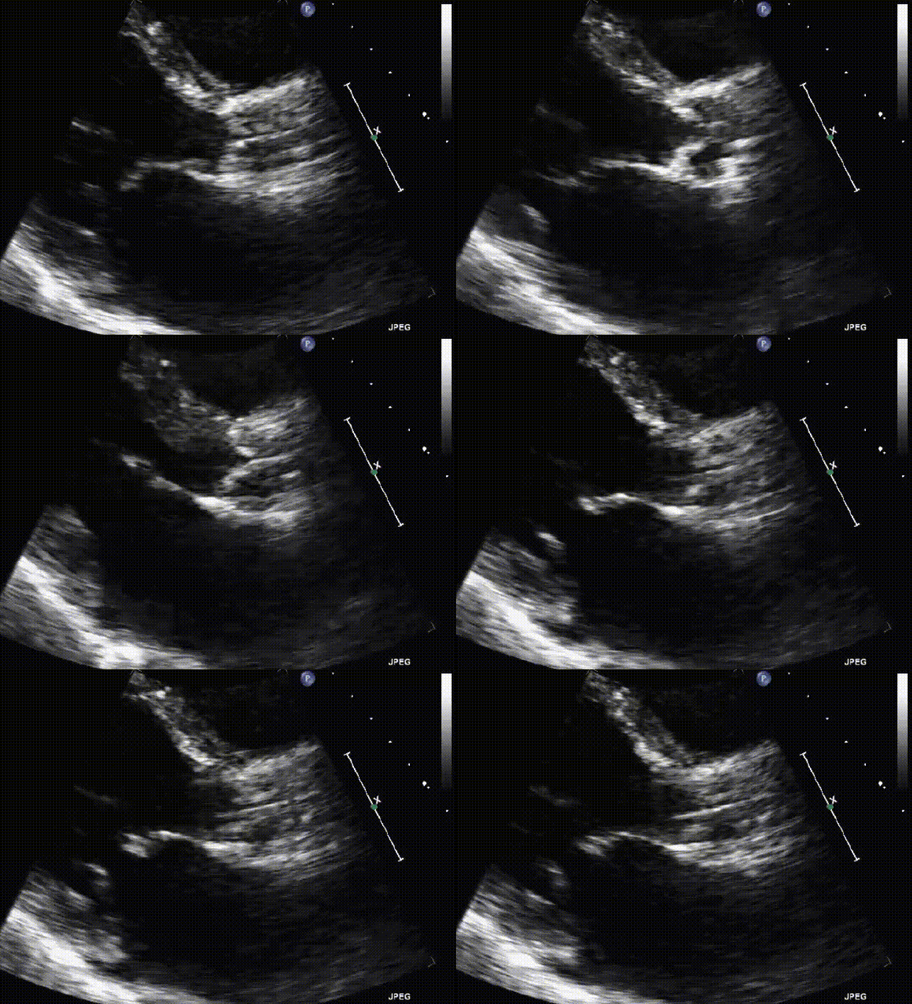
Aortic Valve Disease
- Stenosis features:
- Thickened, calcified leaflets
- Restricted opening
- Left ventricular hypertrophy
- Post-stenotic dilation of ascending aorta
- Regurgitation features:
- Incomplete leaflet coaptation
- LV dilation
- Hyperdynamic LV in chronic cases
Mitral Valve Disease
- Stenosis features:
- Thickened, calcified leaflets
- Restricted opening (“hockey stick” appearance)
- Left atrial enlargement
- Normal LV size in pure stenosis
- Regurgitation features:
- Incomplete leaflet coaptation
- Flail or prolapsed leaflet
- Left atrial and ventricular enlargement
- Hyperdynamic LV in chronic cases
Confirmation Steps
MULTI-VIEW CONFIRMATION
- Pericardial effusion:
- Confirm in at least two views
- Differentiate from pleural effusion
- Verify location (circumferential vs. loculated)
- Cardiomyopathies:
- Assess in multiple views for comprehensive evaluation
- Measure chamber dimensions when possible
- Evaluate both structure and function
- Valvular disease:
- Assess valve in multiple views
- Look for chamber remodeling responses
- Correlate findings between views
CLINICAL CORRELATION
- Critical integration:
- Combine POCUS findings with clinical presentation
- Assess hemodynamic impact
- Consider pre-existing conditions
- Intervention guidance:
- Identify findings requiring immediate action
- Determine severity and urgency
- Guide specific therapeutic approaches
- Follow-up considerations:
- Recommend formal echocardiography when appropriate
- Consider serial POCUS examinations
- Monitor response to interventions
Documentation Requirements
- Specific pathologic findings documented with images
- Measurements where applicable
- Integration with clinical context
- Presence and severity of key pathologies
- Views in which abnormalities were identified
- Hemodynamic impact assessment
- Recommendations for further evaluation
SCOPE GUIDE
Strategies & Clinical Optimization
Pericardial Effusion Assessment
- View optimization
- Examine all views as distribution may be variable
- Subcostal view often best for anterior effusions
- Adjust depth to visualize entire pericardium
- Tamponade recognition
- Look specifically for chamber collapse and IVC plethora
- RA collapse most sensitive (earliest sign)
- RV collapse more specific
- Effusion characteristics
- May first appear posteriorly in PLAX view
- Circumferential effusions more likely significant
- Dark, echo-free space suggests simple fluid
- Echogenic effusions suggest blood, exudate, or infection
Cardiomyopathy Recognition
- Dilated cardiomyopathy approach
- Measure chamber dimensions when possible
- Assess for spherical remodeling
- Evaluate for secondary mitral regurgitation
- Hypertrophic cardiomyopathy approach
- Measure septal and posterior wall thickness
- Look for systolic anterior motion of mitral valve
- Assess for dynamic outflow obstruction
- Restrictive patterns
- Normal-sized ventricles with biatrial enlargement
- Evaluate for pericardial thickening
- IVC dilation with reduced respiratory variation
Valvular Assessment Optimization
- Aortic valve approach
- PLAX and PSAX views most useful
- Assess leaflet thickness and mobility
- Evaluate for secondary chamber changes
- Mitral valve approach
- PLAX and apical views most useful
- Assess for leaflet thickening, restriction, or prolapse
- Evaluate LA and LV size responses
- Right-sided valves
- Apical and subcostal views most useful
- Assess for tricuspid regurgitation signs
- Look for RV and RA size responses
Pearls
- Not all effusions cause tamponade; clinical correlation is essential
- Small, loculated effusions can cause tamponade if rapid accumulation
- In tension pneumothorax, look for mediastinal shift and RV compression
- Cardiac findings should not be interpreted in isolation from clinical context
- Serial examinations provide valuable trending information
Pathology-Specific Tips
- Pericardial effusion pearls:
- Effusion is seen anterior to descending aorta (vs. pleural effusion)
- Small effusions may be physiologic (women, post-surgery)
- Tamponade is a clinical diagnosis supported by POCUS
- Cardiomyopathy pearls:
- Regional wall motion abnormalities suggest ischemia
- Global dysfunction suggests non-ischemic etiology
- LV thrombus risk increases with severe dysfunction
- Valvular pearls:
- Combined stenosis and regurgitation are common
- Acute regurgitation has different appearance than chronic
- Functional regurgitation results from chamber dilation
Quick Resources
Effusion Size Classification
(diagram)
Key Measurements
- Effusion size classification:
- Small: <1.0 cm
- Moderate: 1.0-2.0 cm
- Large: >2.0 cm
- LV hypertrophy: Wall thickness >1.1 cm
- Dilated LV: End-diastolic diameter >5.6 cm
- Dilated LA: Diameter >4.0 cm
Key Images/Diagrams

Pathology Recognition
- Effusion localization patterns
- Tamponade physiology illustration
- Cardiomyopathy comparison chart
- Valvular disease patterns
Management Guidance
- Tamponade recognition checklist
- Cardiomyopathy pattern reference
- Valvular pathology decision tree
- Emergency intervention indicators
Reference Materials
- Effusion vs. pleural fluid differentiation
- Tamponade demonstration images
- Cardiomyopathy comparison examples
- Valvular abnormality reference images
References
1. International consensus guidelines on cardiac POCUS
2. American Society of Echocardiography guidelines for chamber quantification
3. American College of Emergency Physicians. ACEP Policy Statement: Emergency Ultrasound Imaging Criteria Compendium
Media Attributions
- PLAX aortic stenosis is licensed under a CC BY-NC (Attribution NonCommercial) license

